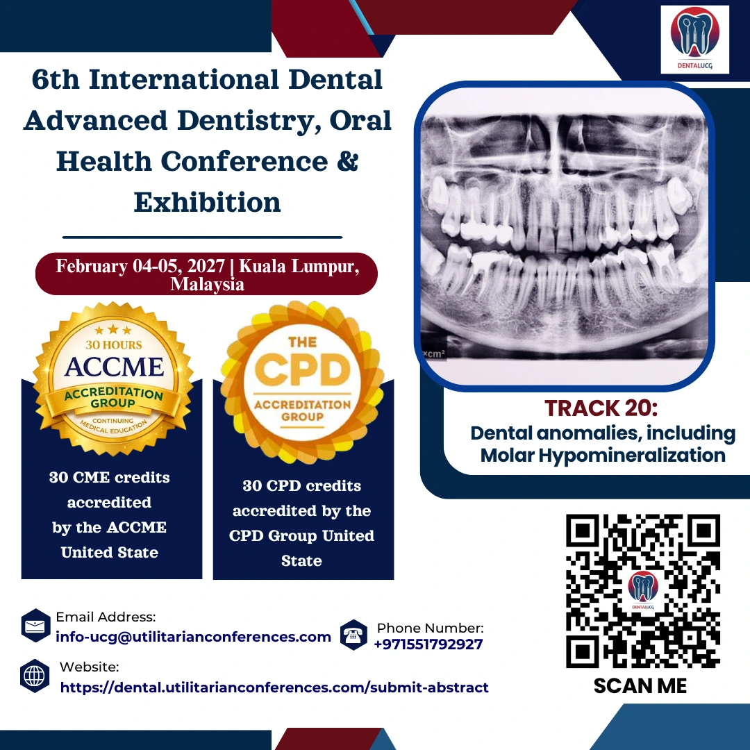
Dental Anomalies and Molar Hypo mineralization: Identification, Etiology, and Management
Dental anomalies are variations from normal tooth development that may affect the number, size, shape, structure, or position of teeth. These conditions can arise from genetic factors or environmental influences and often require early diagnosis and individualized management to maintain both function and aesthetics.
Common Types of Dental Anomalies
- Hypodontia: Congenital absence of one or more teeth
- Hypodontia: Presence of supernumerary (extra) teeth
- Amelogenesis Imperfecta: Inherited enamel formation disorder
- Dentinogenetic Imperfecta: Genetic condition affecting dentin structure
- Paurodontids: Enlarged pulp chambers, commonly in molars
- Fusion and Gemination: Abnormal union or duplication of developing teeth
- Microdontia / Macrodontia: Abnormally small or large teeth
- Peg-shaped Lateral Incisors: Narrow, conical-shaped incisors
- Ectopic Eruption: Teeth erupting in atypical positions
- Transposition: Exchange of positions between adjacent teeth
Molar Hypo mineralization (MIH)
What is MIH?
Molar-Incisor Hypo mineralization (MIH) is a developmental enamel defect of systemic origin that primarily affects first permanent molars and often incisors. The enamel in affected teeth is weak and porous, making it susceptible to breakdown, sensitivity, and rapid caries progression.
Clinical Features of MIH
-
Well-defined white, yellow, or brown enamel opacities
-
Post-eruptive enamel breakdown (PEB)
-
Increased sensitivity to thermal stimuli and brushing
-
Accelerated development of dental caries
-
Challenges in achieving effective local anesthesia
-
Possible behavioral concerns due to chronic discomfort
Causes of MIH
-
Environmental disturbances during early enamel formation (birth to 3 years)
-
Childhood illnesses, especially fever and respiratory infections
-
Use of certain antibiotics (e.g., amoxicillin)
-
Prenatal influences such as maternal illness or nutritional deficiencies
-
Potential genetic predisposition
Diagnosis
Diagnosis is primarily clinical and based on:
-
Presence of demarcated enamel opacities
-
Evidence of post-eruptive enamel loss
-
Atypical restorations in first permanent molars
-
Consideration of extraction in severe cases, typically between ages 8–10